Functional MRI sheds new light on AE
September 9, 2018
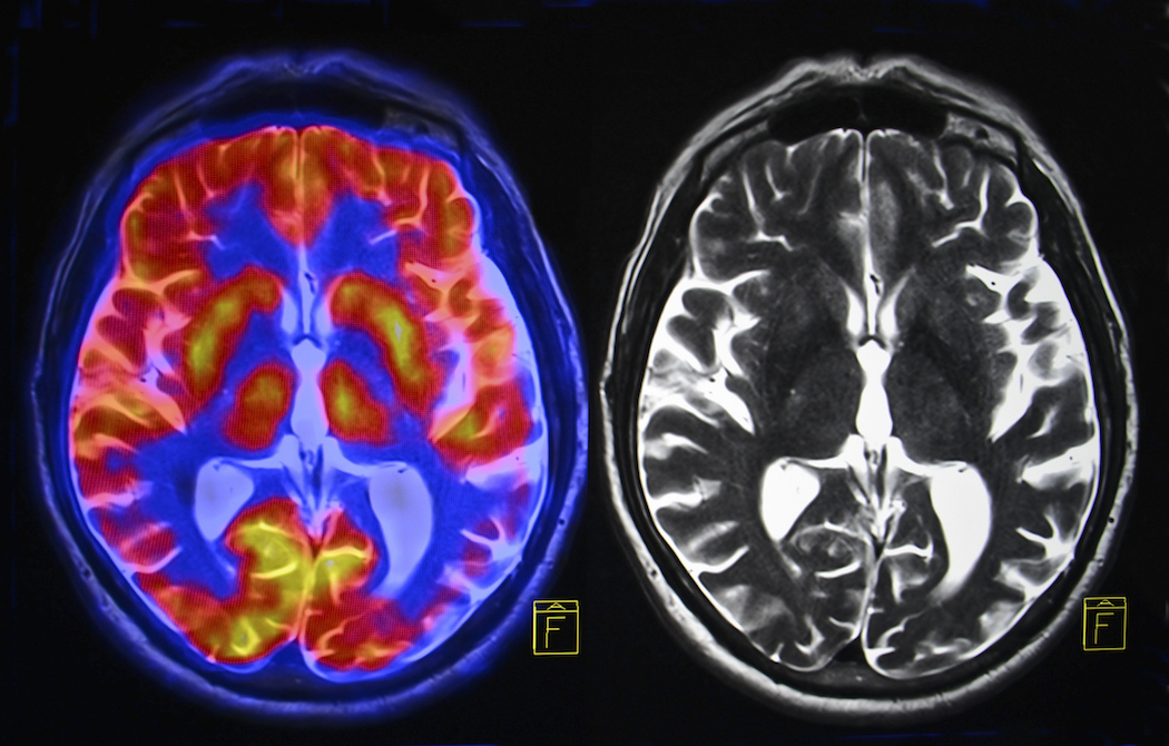
New methods of studying the brain have been emerging in recent decades. One such method is the use of functional MRI, which is a way to measure how the brain works as opposed to regular MRI, which looks at structure. This week an exciting new research study was released by the Autoimmune Neurology Group in Oxford, UK. This was the first study to use functional MRI to measure the brain activity of AE patients with the anti-LGI1 antibody who experience faciobrachial seizures.
Eight AE patients in the recovery phase participated in the study, along with eight healthy volunteers as a comparison. They were asked to view familiar or unfamiliar scenes while images of their brain were scanned. Researchers were particularly focused on the hippocampus. This is the part of the brain often affected in AE and responsible in part for memory, emotions and other functions. Patients often develop lingering cognitive difficulties after AE, mostly with memory. Evidence has pointed to structural damage in the hippocampus as a potential cause for these difficulties, however the authors wanted to explore whether functional aspects of the brain may be contributing in some cases.
They found that indeed, many of those who had previously been affected by AE showed abnormal hippocampal activity (or function), while the healthy volunteers did not. They stated that this may be due to the frequent seizures experienced previously during the illness. The authors mentioned that these results may have implications for future treatment methods to lessen long-term cognitive difficulties in AE.
The full article can be read here.